Introduction
Injury or accidents can affect any structure of the eyelid, eyeball and the surrounding bones (fracture). Laceration of the eyelid needs meticulous placement of stitches to preserve its normal shape and function. Injury to the tear ducts (canaliculus) within the eyelids require special placement of silicone tubes within them to keep them open while the injured eyelid heals. A thorough examination is also done to find out if the eyeball is also injured.
Fractures of the bone surrounding the eye (orbital walls) may cause the eye to sink back into its socket, lead to a facial deformity, poor eye movement, or loss of vision. A clinical examination combined with a CT scan of the orbit can easily detect any of this possibility. The presence of any retained foreign bodies also needs to be ruled out. Orbital fractures can now be repaired with minimal incisions through the inside of the eyelid or mouth. Most often, a barrier has to be placed to cover the fracture site. Excellent synthetic plates (polyethylene or silastic) are available for this purpose, or alternatively a small sliver of patient’s own bone can be safely removed from elsewhere and placed over the fracture site.
Surgical time and recovery
Eyelid laceration repair can be performed under local or general anesthesia, depending upon the location and extent of the injury. Fracture repair almost always requires general anesthesia. Trauma repair can take anything from 30 minutes to 3 hours.
Discomfort is controlled with medications after surgery, and lasts for a week. Stitches are removed in 5-7 days. Most patients are back to light work within a week.
Insurance
Health insurance will cover the costs of trauma repair.
BEFORE AND AFTER PHOTOS
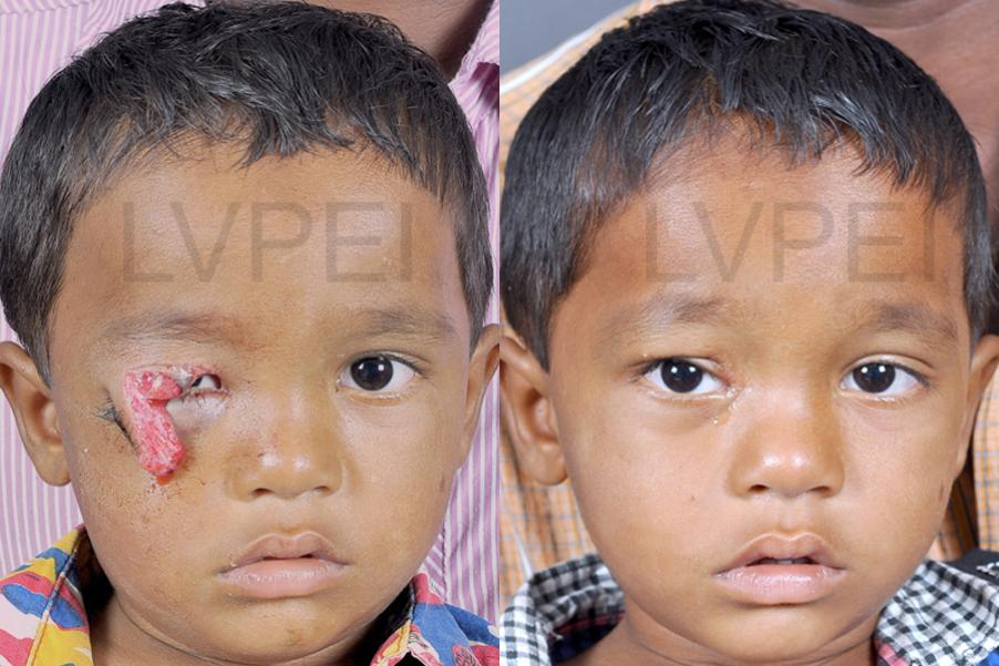
A 4-year-old boy with avulsion injury to his eyelid. Meticulous stepwise repair of each eyelid component can bring back the appearance to near-normal, while preserving safety of the eye.
An unfortunate dob-bite injury leaves this child's eyelids into pieces. A complex surgery to place back every functional component of the eyelid gives a good cosmetic and functional result (A minor upper eyelid droop remains, but it can be further improved at a later date).

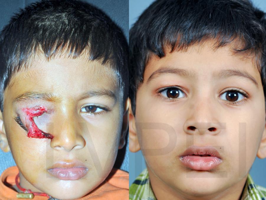
Another avulsion injury of the upper eyelid, treated with reconstructive plastic surgery.
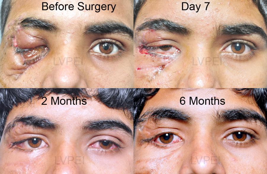
Injury around the eyelids with loss of tissue. First, anti-scarring injections are used, followed by an eyelid-plastic surgery to bring it back to normal appearance.
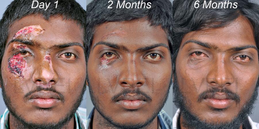
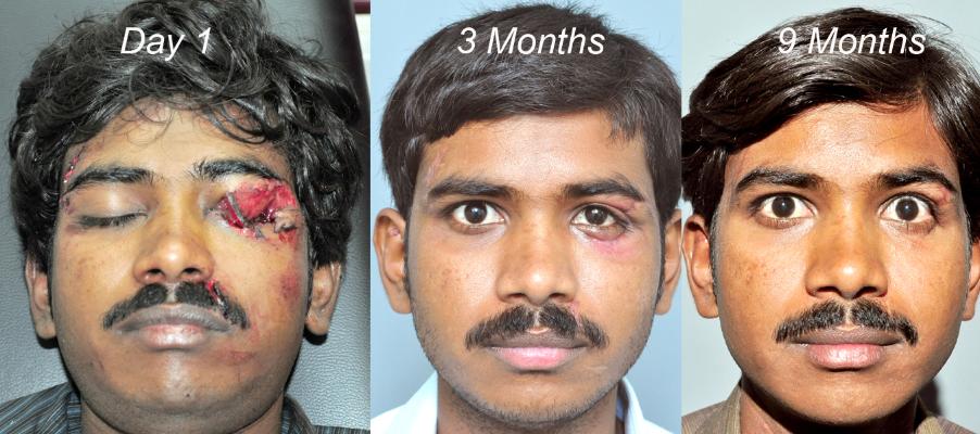
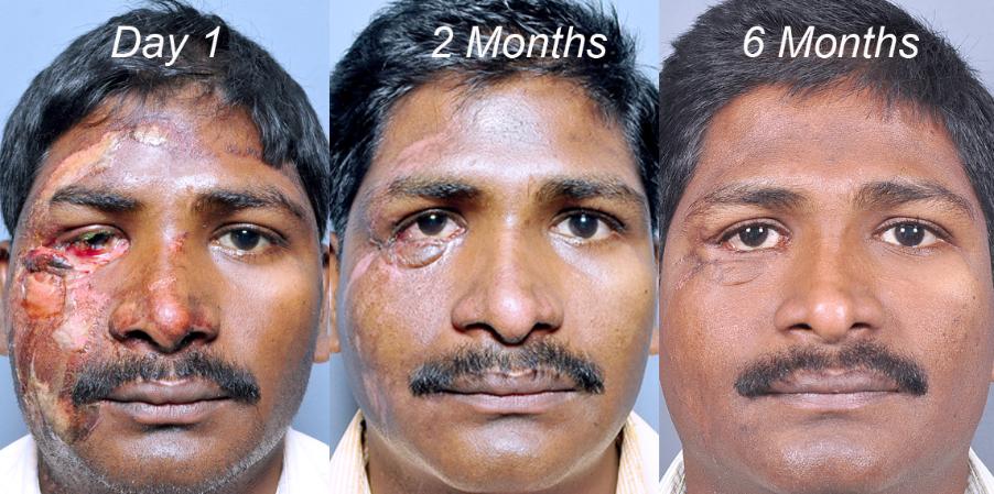
Blast injury leading to foreign bodies, and torn eyelids. Staged plastic reconstruction of the eyelids has restored a near-normal appearance in few months.
Road traffic accident leading to loss of entire lower eyelid. A series of reconstructive surgeries has improved the appearance and protected the eyeball (Always wear a helmet!).
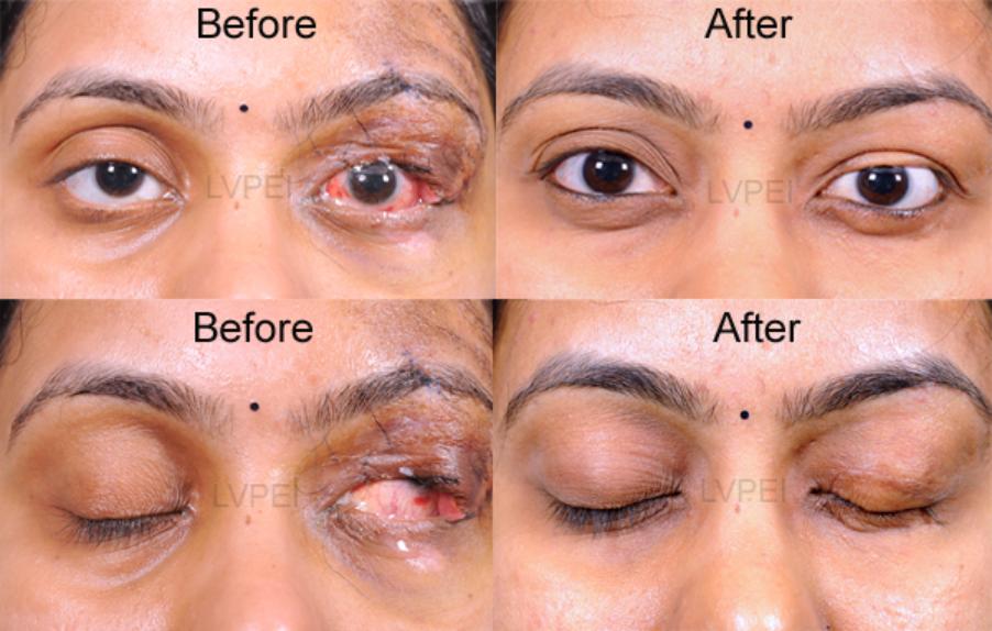
Left upper eyelid was lost after injury, exposing the eyeball. A two stage eyelid reconstruction (Cutler-Beard procedure) helped restore the normal eyelid structures.